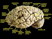
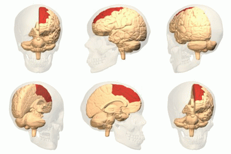
Category:Gyri
Saltar para a navegação
Saltar para a pesquisa
saliência na superfície do cérebro, cercado por um ou mais sulcos | |||||
| Carregar ficheiro | |||||
| Instância de |
| ||||
|---|---|---|---|---|---|
| Subclasse de |
| ||||
| Parte de | |||||
| |||||

see also
and
Subcategorias
Esta categoria contém as seguintes 28 subcategorias (de um total de 28).
- SVG gyri (2 F)
A
- Angular gyrus (29 F)
C
- Cuneus (22 F)
F
G
H
- Heschl's gyrus (32 F)
I
- Inferior temporal gyrus (30 F)
L
- Lateral occipital gyrus (9 F)
M
- Middle frontal gyrus (32 F)
- Middle temporal gyrus (25 F)
O
P
- Paracentral lobule (22 F)
- Planum temporale (15 F)
- Postcentral gyrus (42 F)
- Precentral gyrus (40 F)
- Precuneus (33 F)
S
- Straight gyrus (18 F)
- Subcallosal area (5 F)
- Superior frontal gyrus (44 F)
- Superior parietal lobule (18 F)
- Supramarginal gyrus (28 F)
Multimédia na categoria "Gyri"
Esta categoria contém os seguintes 200 ficheiros (de um total de 204).
(página anterior) (página seguinte)-
"Traite complet de l'anatomie...",Foville, 1844 Wellcome L0019133.jpg 1 230 × 1 557; 552 kB
-
"Traite complet de l'anatomie...",Foville, 1844 Wellcome L0019135.jpg 3 763 × 4 947; 4,58 MB
-
04 2 facies ventralis cerebri gyri.jpg 1 799 × 2 122; 644 kB
-
A human head dissected; "In memoriam". Wellcome L0074866.jpg 4 817 × 5 611; 4,43 MB
-
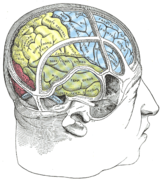
Brain parcellation.jpg 600 × 320; 81 kB
-
Brain surface by Raymond Vieussens, 1684.png 455 × 386; 220 kB
-
Brain Surface Gyri.SVG 1 024 × 731; 28 kB
-
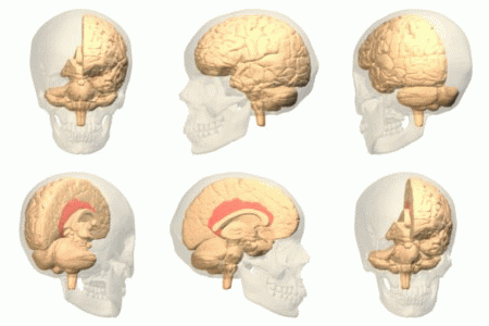
Brain visualization.jpg 500 × 500; 40 kB
-
Brain-1.jpg 484 × 356; 58 kB
-
Brain-disease-gyrification.png 1 794 × 550; 620 kB
-
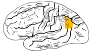
Brodmann area 3 1 2.png 300 × 190; 20 kB
-
Causes of Autism in Brain.png 960 × 620; 194 kB
-
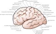
Cerebral cortex, side view.svg 2 395 × 1 449; 137 kB
-
Cerebral Gyri - Inferior Surface2.png 757 × 569; 431 kB
-
Cerebral Gyri - Insula.png 761 × 571; 293 kB
-
Cerebral Gyri - Lateral Surface.png 728 × 546; 320 kB
-
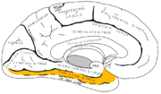
Cerebral Gyri - Medial Surface1.png 730 × 547; 366 kB
-
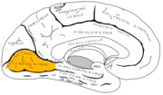
Cerebral Gyri - Medial Surface2.png 727 × 547; 301 kB
-
Cingulate gyrus animation small.gif 150 × 150; 470 kB
-
Constudproc.png 763 × 865; 31 kB
-
Coronal hippocampe.png 543 × 499; 298 kB
-
Coronal insula.png 560 × 409; 147 kB
-
CorpusCallosum.svg 1 025 × 598; 25 kB
-
Cunningham cerebral sulci.png 1 252 × 790; 2,84 MB
-
Desikan-Killiany atlas regions.pdf 3 150 × 1 918; 13,59 MB
-
Destrieux atlas regions.pdf 5 864 × 2 850; 28,77 MB
-
EB1911 Brain Fig. 11-Gyri and Sulci on Mesial aspect.jpg 682 × 393; 45 kB
-
EB1911 Brain Fig. 9-Gyri and Sulci.jpg 672 × 412; 57 kB
-
F. Vicq d'Azyr, Planches pour le traite de Wellcome L0000998.jpg 3 322 × 3 178; 5,03 MB
-
Face sup T1.png 563 × 482; 222 kB
-
Facies ventralis cerebri.jpg 1 834 × 2 068; 629 kB
-
File-Cerebral Gyri - Inferior Surface1.png 761 × 569; 419 kB
-
Frontal gyrus coronal sections.gif 148 × 158; 1,24 MB
-
Frontal gyrus sagittal sections.gif 185 × 158; 1,41 MB
-
Frontal gyrus transversal sections.gif 148 × 185; 1,39 MB
-
FrontalCapts.png 2 309 × 911; 927 kB
-
Gehirn Frontalschnitt hippocampus-it.png 1 055 × 573; 247 kB
-
Gehirn Frontalschnitt hippocampus.png 913 × 573; 263 kB
-
Gehirn lateral gyri el.svg 669 × 519; 67 kB
-
Gehirn lobi medial.png 819 × 598; 61 kB
-
Gehirn, lateral - Hauptgyri + Hauptsulci.svg 624 × 498; 56 kB
-
Gehirn, lateral - Hauptgyri beschriftet.svg 624 × 498; 334 kB
-
Gray1197.png 444 × 500; 47 kB
-
Gray725 anterior central gyrus.png 255 × 600; 61 kB
-
Gray725 cant been seen top.png 663 × 1 560; 205 kB
-
Gray725 frontal pole.png 255 × 600; 57 kB
-
Gray725 inferior frontal gyrus.png 255 × 600; 60 kB
-
Gray725 middle frontal gyrus.png 255 × 600; 61 kB
-
Gray725 occipital pole.png 255 × 600; 57 kB
-
Gray725 posterior central gyrus.png 255 × 600; 61 kB
-
Gray725 superior frontal gyrus.png 255 × 600; 61 kB
-
Gray725 superior parietal lobule.png 255 × 600; 60 kB
-
Gray725.png 255 × 600; 18 kB
-
Gray726 angular gyrus.png 992 × 573; 178 kB
-
Gray726 ar.svg 992 × 573; 409 kB
-
Gray726 cant been seen lateral.png 842 × 487; 177 kB
-
Gray726 frontal pole.png 992 × 573; 174 kB
-
Gray726 inferior frontal gyrus.png 992 × 573; 177 kB
-
Gray726 inferior parietal lobule (hy).png 2 108 × 1 217; 909 kB
-
Gray726 inferior parietal lobule.png 992 × 573; 182 kB
-
Gray726 inferior temporal gyrus.png 992 × 573; 169 kB
-
Gray726 middle frontal gyrus.png 992 × 573; 192 kB
-
Gray726 middle temporal gyrus.png 992 × 573; 173 kB
-
Gray726 occipital pole.png 992 × 573; 170 kB
-
Gray726 opecular part of IFG.png 992 × 573; 172 kB
-
Gray726 orbital part of IFG.png 992 × 573; 170 kB
-
Gray726 postcentral gyrus.png 992 × 573; 117 kB
-
Gray726 precentral gyrus.png 992 × 573; 181 kB
-
Gray726 superior frontal gyrus.png 992 × 573; 179 kB
-
Gray726 superior parietal lobule.png 992 × 573; 174 kB
-
Gray726 superior temporal gyrus.png 992 × 573; 173 kB
-
Gray726 supramarginal gyrus.png 992 × 573; 182 kB
-
Gray726 temporal pole.png 992 × 573; 170 kB
-
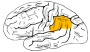
Gray726 triangular part of IFG.png 992 × 573; 174 kB
-
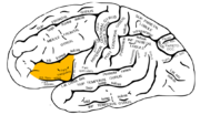
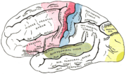
Gray726.png 700 × 405; 29 kB
-
Gray726.svg 992 × 573; 146 kB
-
Gray727 anterior cingulate cortex.png 1 025 × 598; 141 kB
-
Gray727 cant been seen medial.png 870 × 507; 147 kB
-
Gray727 cingulate gyrus.png 1 025 × 598; 92 kB
-
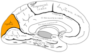
Gray727 Cuneus.png 1 025 × 598; 144 kB
-
Gray727 frontal pole.png 1 025 × 598; 137 kB
-
Gray727 fusiform gyrus.png 1 025 × 598; 135 kB
-
Gray727 inferior temporal gyrus.png 1 025 × 598; 134 kB
-
Gray727 latin.svg 1 025 × 598; 23 kB
-
Gray727 lingual gyrus.png 1 025 × 598; 134 kB
-
Gray727 occipital pole.png 1 025 × 598; 134 kB
-
Gray727 paracentral gyrus.png 1 025 × 598; 136 kB
-
Gray727 parahippocampal gyrus.png 1 025 × 598; 134 kB
-
Gray727 precuneus.png 1 025 × 598; 143 kB
-
Gray727 superior frontal gyrus.png 1 025 × 598; 142 kB
-
Gray727 temporal pole.png 1 025 × 598; 134 kB
-
Gray727 uncus of parahippocampal gyrus.png 1 025 × 598; 133 kB
-
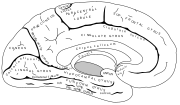
Gray727.svg 1 025 × 598; 18 kB
-
Gray729 frontal pole.png 300 × 366; 60 kB
-
Gray729 orbital gyrus.png 300 × 366; 59 kB
-
Gray729 straight gyrus.png 300 × 366; 61 kB
-
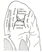
Gray729.png 300 × 366; 20 kB
-
Gray743 cingulate gyrus.png 450 × 542; 60 kB
-
Gray743 inferior frontal gyrus.png 450 × 542; 172 kB
-
Gray743 insular cortex.png 450 × 542; 170 kB
-
Gray743 middle frontal gyrus.png 450 × 542; 171 kB
-
Gray743 straight gyrus.png 450 × 542; 170 kB
-
Gray743 superior frontal gyrus.png 450 × 542; 171 kB
-
Gray756.png 600 × 354; 32 kB
-
Gray757.png 600 × 346; 27 kB
-
Gyri and sulci of frontal cortex of monkey brain (Cebus apella).jpg 1 200 × 592; 99 kB
-
Gyri Basal no text1.png 572 × 719; 595 kB
-
Gyri Basal no text2.png 567 × 690; 584 kB
-
Gyri Insula no text.png 697 × 524; 399 kB
-
Gyri Lateral no text.png 632 × 484; 413 kB
-
Gyri Medial no text.png 644 × 498; 552 kB
-
Gyri Medial no text2.png 647 × 505; 444 kB
-
Gyrus cinguli.png 800 × 467; 116 kB
-
Gyrus externe droit.png 1 100 × 800; 261 kB
-
Gyrus externe.png 666 × 450; 112 kB
-
Gyrus sulcus ja.png 960 × 720; 115 kB
-
Gyrus sulcus.png 960 × 720; 95 kB
-
Hand-book of physiology (1892) (14578661130).jpg 1 796 × 1 516; 282 kB
-
Hand-book of physiology (1892) (14578725808).jpg 1 164 × 1 476; 185 kB
-
Hand-book of physiology (1892) (14785228633).jpg 1 776 × 1 128; 204 kB
-
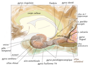
Hippocampe parahippo.png 1 000 × 800; 426 kB
-
Homunculus-ja paracentral lobule.png 1 125 × 578; 189 kB
-
Human and chimp brain.png 1 010 × 1 346; 836 kB
-
Human brain inferior view description 2.JPG 373 × 467; 36 kB
-
Human brain inferior-medial view description 2.JPG 702 × 491; 68 kB
-
Human brain inferior-medial view description 3.JPG 702 × 491; 63 kB
-
Human brain inferior-medial view with marked Precuneus.JPG 702 × 491; 211 kB
-
Human brainstem anterior view 2 description.JPG 346 × 487; 35 kB
-
Human Cortical Development.png 2 303 × 2 656; 1 022 kB
-
Inferieur gyrus.png 724 × 652; 207 kB
-
Inferior frontal gyrus animation small.gif 150 × 150; 570 kB
-
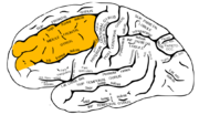
Inferior frontal gyrus.png 300 × 190; 40 kB
-
Infero interne gyrus.png 579 × 449; 106 kB
-
Lateral surface - Inferior frontal gyrus.png 1 236 × 800; 597 kB
-
Lateral surface - Middle frontal gyrus.png 1 236 × 800; 596 kB
-
Lateral surface - opercular part of the inferior frontal gyrus.png 1 236 × 800; 593 kB
-
Lateral surface - Orbital part of inferior frontal gyrus.png 1 236 × 800; 594 kB
-
Lateral surface - Superior frontal gyrus.png 1 236 × 800; 594 kB
-
Lateral surface - triangular part of inferior frontal gyrus.png 1 236 × 800; 593 kB
-
Lateral surface of cerebral cortex - gyri.png 1 236 × 800; 760 kB
-
Lissencephaly.jpg 490 × 315; 17 kB
-
Lk444.jpg 500 × 667; 70 kB
-
Macaque monkey's premotor areas.jpg 1 200 × 1 108; 156 kB
-
Man&chimpbrains.png 591 × 558; 455 kB
-
Medial paracentral lob.png 673 × 481; 130 kB
-
Medial surface - Sperior frontal gyrus.png 1 179 × 747; 515 kB
-
Medial surface of cerebral cortex - ceneus.png 1 179 × 747; 514 kB
-
Medial surface of cerebral cortex - entorhinal cortex.png 1 179 × 747; 515 kB
-
Medial surface of cerebral cortex - fusiform gyrus.png 1 179 × 747; 515 kB
-
Medial surface of cerebral cortex - gyri.png 1 179 × 747; 644 kB
-
Medial surface of cerebral cortex - lingual gyrus.png 1 179 × 747; 515 kB
-
Medial surface of cerebral cortex - parahippocampal.png 1 179 × 747; 515 kB
-
Medial surface of cerebral cortex - preceneus.png 1 179 × 747; 514 kB
-
Middle frontal gyrus.png 300 × 190; 42 kB
-
OccCapts.png 1 662 × 781; 847 kB
-
Operculum.png 700 × 405; 59 kB
-
Operculum1.jpg 1 065 × 796; 146 kB
-
Orbital gyrus viewed from bottom.png 504 × 637; 268 kB
-
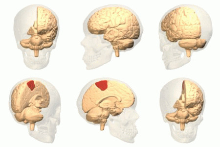
Paracentral lobule animation small.gif 150 × 150; 568 kB
-
Parahippocampe.png 1 032 × 761; 622 kB
-
ParcellationBrains.jpg 364 × 384; 118 kB
-
ParietCapts.png 2 337 × 878; 819 kB
-
Postcentral gyrus 3d.png 600 × 600; 177 kB
-
Postcentral gyrus.gif 250 × 250; 1,87 MB
-
Pre- and post-central gyrus, right hemisphere cropped.png 426 × 488; 140 kB
-
Pre- and post-central gyrus, right hemisphere.jpg 884 × 1 394; 1,13 MB
-
Precentral gyrus 3d.png 600 × 600; 178 kB
-
Precentral gyrus.jpg 700 × 405; 47 kB
-
Precuneus connectivity new.gif 343 × 447; 30 kB
-
Precuneus connectivity.jpg 4 111 × 6 939; 81,65 MB
-
PretermSurfaces HiRes es.png 876 × 190; 158 kB
-
PretermSurfaces HiRes ja.png 876 × 190; 146 kB
-
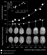
PretermSurfaces HiRes.png 876 × 190; 165 kB
-
PSM V35 D759 Diagram of the left cerebral hemisphere.jpg 1 626 × 1 413; 254 kB
-
Sagittale-insula-Heschl.png 579 × 420; 140 kB
-
Slide2HAN.JPG 960 × 720; 96 kB
-
Slide3HAN.JPG 960 × 720; 117 kB
-
Smith.PNG 1 500 × 928; 735 kB
-
Sobo 1909 623 ar.png 2 404 × 2 652; 5,62 MB
-
Sobo 1909 623.png 2 404 × 2 652; 18,27 MB
-
Sobo 1909 624.png 3 060 × 2 247; 19,71 MB
-
Sobo 1909 625.png 1 137 × 703; 2,29 MB
-
Sobo 1909 626.png 1 326 × 899; 3,42 MB
-
Sobo 1909 627.png 983 × 981; 2,76 MB
-
Sobo 1909 628.png 1 058 × 1 159; 3,52 MB
-
Sobo 1909 629.png 991 × 1 015; 98 kB
-
Sobo 1909 630.png 1 077 × 1 239; 3,83 MB
-
Sobo 1909 631.png 1 010 × 689; 1,99 MB
-
Sobo 1909 632.png 1 363 × 879; 3,43 MB
-
Sobo 1909 633.png 1 078 × 699; 2,16 MB
-
Sobo 1909 634.png 1 089 × 703; 2,19 MB
-
Some brain areas.png 989 × 412; 146 kB
-
Standard anatomical parcellation of the posterior cortical surface.png 1 810 × 1 717; 1,26 MB
-
Straight gyrus - inferior view.png 500 × 638; 284 kB
-
Straight gyrus animation.gif 300 × 300; 1,81 MB
-
Superieur gyrus.png 554 × 516; 115 kB
-
Superior frontal gyrus animation small.gif 150 × 150; 549 kB
-
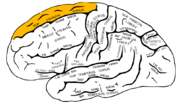
Superior frontal gyrus.png 300 × 190; 32 kB
-
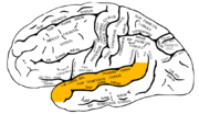
Superior temporal gyrus.png 300 × 190; 35 kB


